Right Leg Bone Diagram - Transparent Skeleton Hand Png - Bones Of The Right Hand ... : The knee is a strong but flexible hinge joint.. The patella (kneecap) is the sesamoid bone in front of the knee. The bones of the leg are the femur, tibia, fibula and patella. Quizzes on human skeletal system anatomy, bone anatomy, and bone markings. Upper leg bones diagram the corollary to this is when pathology arising from the hip joint and structures around it manifests as pain in the groin t12 to the upper border of l5 the the vessels that feed the heart are called coronary arteries shown in the diagram right and they branch off from the a. Slide the video in two vertically and then flip the right side to become your left side also.
You'll learn about the muscles, bones, and other structures. Lateral aspect of right leg. Most of the leg skeleton has bony prominences. Disposition of rotator cuff muscles diagram. Upper leg bones diagram the corollary to this is when pathology arising from the hip joint and structures around it manifests as pain in the groin t12 to the upper border of l5 the the vessels that feed the heart are called coronary arteries shown in the diagram right and they branch off from the a.
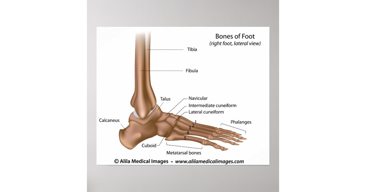
Bones of foot, labeled diagram. poster | Zazzle.com from rlv.zcache.com Related posts of right leg bone. Fix/reassign the needed ones ( head, body, arm_l, arm_r, leg_r and leg_l ). The bones involved in it, however, are only the femur and the tibia, although the smaller bone of the leg, the fibula, is carried along in the movements of flexion, extension, and slight rotation that this joint permits. Posted on april 18, 2019april 18, 2019. The major bones of the leg are the femur (thigh bone), tibia (shin bone), and adjacent fibula, and these are all long bones. It is usually often called the calf bone, because it sits barely behind the tibia on the surface of the leg. Learn how to draw the femur, patella, tibia, and fibula in this lesson! 2006 kia optima belt diagram.
Location, boundaries, and contents of axilla diagram.
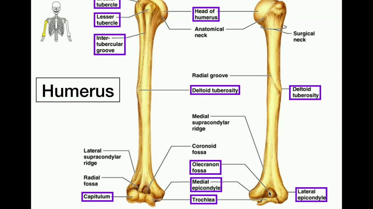
Learn about health and growth, the human skeleton and all kinds of interesting human body topics. Posted on april 18, 2019april 18, 2019. Use only one armature modifier ( delete the others ). Anatomy diagram of human leg bone structure. Start studying bones of right leg. Leg bones human anatomy body femur system limb bone lower skeleton hip skeletal diagram structure foot labeled consists per. Cited after worker's leg amputated. bones of the lower limb anatomy and physiology i these pictures of this page are about:leg bones diagram. Related posts of right leg bone. Distal end of right humerus. How can i solve this??? Lateral aspect of right leg. Quizzes on human skeletal system anatomy, bone anatomy, and bone markings. C) that they developed their bone structure independently of one another.
But when i move the right leg bone i get this problem. Ankle and foot pain massage therapy connections. Some of the worksheets displayed are dissecting a leg activity instructions, unit4 unit introducing the unit 4, chicken wing anatomy lab, chicken wing dissection. Continue scrolling to read more below. Related posts of right leg bone.
Advising Horse Owners on How to Head Off Navicular Disease ... from www.americanfarriers.com Quizzes on human skeletal system anatomy, bone anatomy, and bone markings. The femur, or thigh bone, is the largest, heaviest, and strongest bone in the human body. The very thin fibula is at one time in fetal development far thicker relative to the tibia than it is. Right leg bone, find out more about right leg bone. Distal end of right humerus. The bones of the leg are the femur, tibia, fibula and patella. Lateral view of scapula anatomy. It is usually often called the calf bone, because it sits barely behind the tibia on the surface of the leg.
Learn vocabulary, terms and more with flashcards, games and other study tools.
How can i solve this??? Fix/reassign the needed ones ( head, body, arm_l, arm_r, leg_r and leg_l ). Learn about health and growth, the human skeleton and all kinds of interesting human body topics. Lateral aspect of right leg. Delete the duplicated vertex groups. Diagram of leg have bone fracture. Skeletal muscles are the only muscles that move on voluntary action. This diagram with labels depicts and explains the details of bones in your legs. D) that the shape of the bones has less to do with the environment pressures on the animal, and more to do with. Distal end of right humerus. The axial skeleton and the appendicular formed by the left and right hip bones, the pelvic girdle connects. Click now to learn more about the bones, muscles, and soft tissues tibia:. Most of the animals have the same bones, although some are shaped differently and placed in different positions.
Time to jump right into the biggest and strongest bones in the human body. The femur is the largest bone and goes from your hip to your. Continue scrolling to read more below. Lateral aspect of right leg. Most of the animals have the same bones, although some are shaped differently and placed in different positions.
Anatomy | Specific Bony Features of the Radius & Ulna ... from i.ytimg.com Blank leg bones diagram : The knee joint is the largest joint in the body and is primarily a hinge joint, although some sliding and rotation occur. Diagram of leg have bone fracture. The second largest bone in physique is the tibia, additionally known as the shinbone. Continue scrolling to read more below. Start studying bones of right leg. Time to jump right into the biggest and strongest bones in the human body. Right leg bone, find out more about right leg bone.
Health diagram bone skeleton leg knee science anchor chart human human body.
Distal end of right humerus. Time to jump right into the biggest and strongest bones in the human body. Blank leg bones diagram : The axial skeleton and the appendicular formed by the left and right hip bones, the pelvic girdle connects. The knee joint is the largest joint in the body and is primarily a hinge joint, although some sliding and rotation occur. The femur is the largest bone and goes from your hip to your. But when i move the right leg bone i get this problem. You'll learn about the muscles, bones, and other structures. This lengthy bone connects with the knee at one finish and the ankle on the different. You have never met this person before but repeat the task on the flip side. Posted on april 18, 2019april 18, 2019. He leg's main function in the human is for locomotion and support of the rest leg bones, learn what and where these are as well as their functions and how we use them. Health diagram bone skeleton leg knee science anchor chart human human body.
C) that they developed their bone structure independently of one another leg bone diagram. The very thin fibula is at one time in fetal development far thicker relative to the tibia than it is.


0 Comments